
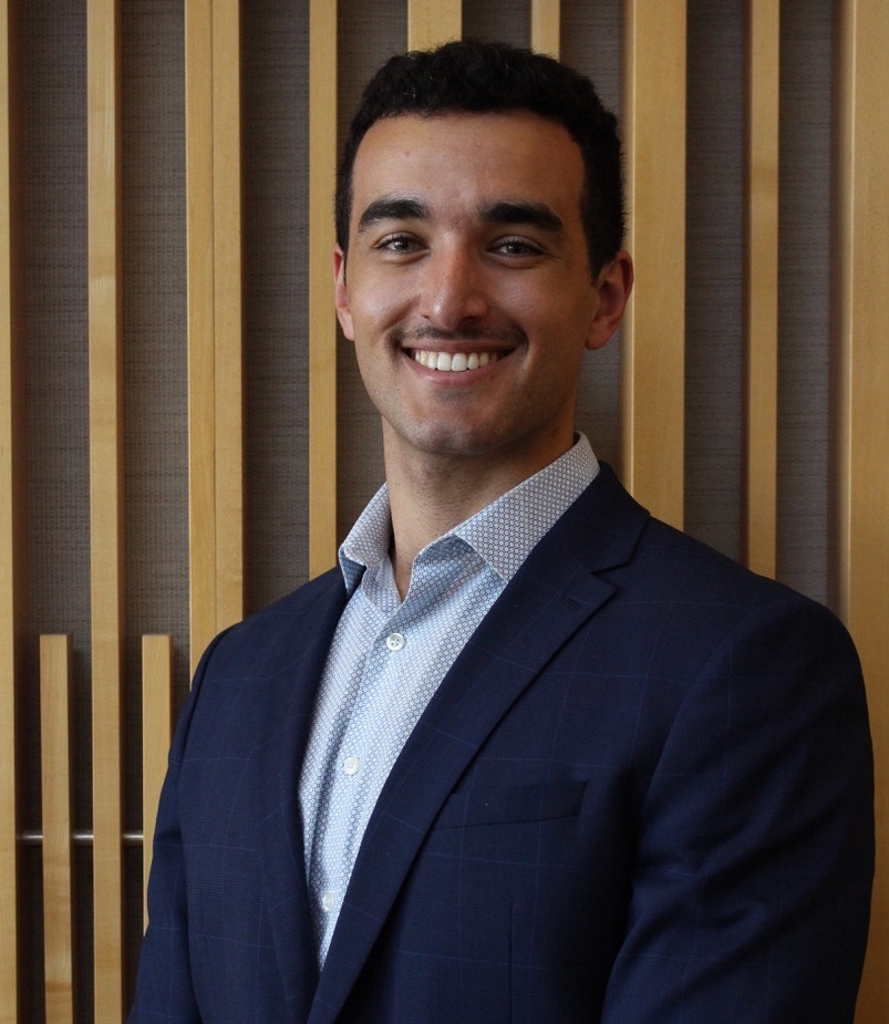
Biography
Robert is a first year MD/PhD student in the Translational Medicine program at Queen’s University. His research investigates the use of artificial intelligence to solve problems related to cancer diagnosis, detection, and differentiation. Previously, he completed both his BSc and MSc in Medical Biophysics from Western University, where he examined the ability to predict treatment response for brain metastases patients and also created learning tools for radiology residents to improve pancreatic cancer detection.
Publications
Maghsoodi, Nooshin; Jamzad, Amoon; Policelli, Robert; Farahmand, Mohammad; Srikanthan, Dilakshan; Kaufmann, Martin; Ren, Kevin Y. M.; Merchant, Shaila; Varma, Sonal; Walker, Ross; McKay, Doug; RUDAN, JOHN; Fichtinger, Gábor; Mousavi, Parvin
Agent-Guided Relational Concept Discovery: Toward Interpretable Surgical Margin Assessment Journal Article
In: arXiv (Cornell University), 2026.
@article{maghsoodi2026,

title = {Agent-Guided Relational Concept Discovery: Toward Interpretable Surgical Margin Assessment},

author = {Nooshin Maghsoodi and Amoon Jamzad and Robert Policelli and Mohammad Farahmand and Dilakshan Srikanthan and Martin Kaufmann and Kevin Y. M. Ren and Shaila Merchant and Sonal Varma and Ross Walker and Doug McKay and JOHN RUDAN and Gábor Fichtinger and Parvin Mousavi},

year  = {2026},

date = {2026-01-01},

journal = {arXiv (Cornell University)},

abstract = {Deep learning models can effectively use Rapid Evaporative Ionization Mass Spectrometry (REIMS) data for surgical margin assessment. However, their clinical adoption remains challenging due to limited generalization to operating room conditions. This difficulty arises because models are typically trained on labeled spectra collected from resected tissue samples, while they must operate on noisy, unlabeled data acquired directly during surgery. In addition, the black-box nature of deep learning models makes it difficult to understand and systematically improve their behavior. Concept-based learning offers a promising way to address these challenges by mapping raw measurements to human-understandable concepts. However, supervised concept-based approaches rely on concept annotations, which are difficult to obtain in complex mass spectrometry workflows. We propose Agent-Guided Concept Discovery, a framework that learns meaningful concepts directly from data without requiring predefined concept labels. During training, a reasoning agent refines semantic descriptions of the learned concepts and adaptively adjusts their weight based on diagnostic relevance. These concepts are further grounded using a biochemical knowledge graph to ensure consistency with known metabolic relationships. Across Skin and Breast Cancer datasets, our model improves balanced accuracy and sensitivity over the baseline. In a representative intraoperative case, it shows fewer false positives, indicating better generalization to surgical conditions.},

keywords = {},

pubstate = {published},

tppubtype = {article}

}
Srikanthan, Dilakshan; Jamzad, Amoon; Wilson, Paul; Maghsoodi, Nooshin; Policelli, Robert; Fichtinger, Gábor; Rudan, John F.; Mousavi, Parvin
Do Foundation Models See Biology? Evaluating Attention Coherence with Spatial Transcriptomics in Glioblastoma Journal Article
In: arXiv (Cornell University), 2026.
@article{srikanthan2026,

title = {Do Foundation Models See Biology? Evaluating Attention Coherence with Spatial Transcriptomics in Glioblastoma},

author = {Dilakshan Srikanthan and Amoon Jamzad and Paul Wilson and Nooshin Maghsoodi and Robert Policelli and Gábor Fichtinger and John F. Rudan and Parvin Mousavi},

year  = {2026},

date = {2026-01-01},

journal = {arXiv (Cornell University)},

abstract = {Whether attention maps from pathology foundation models capture genuine biology remains unknown, yet this question is critical for clinical trust and regulatory approval. We propose a spatial transcriptomics-based framework for orthogonal, hypothesis-free evaluation of attention and apply it to five pathology foundation models (CONCH v1.5, UNI v2, Virchow2, GigaPath, H-Optimus-1) and a ResNet50 baseline. Using attention-based multiple instance learning, we train single-task and multi-task models to predict five molecular alterations in glioblastoma on the CPTAC cohort, validate on an independent TCGA cohort, and evaluate biological coherence of attention maps against 87 transcriptional signatures using co-registered Visium spatial transcriptomics data from 18 samples. Internally, no single encoder dominates across all tasks, and external validation inverts internal performance rankings. Attention maps show a five-fold enrichment gradient from pathways (Cohen's d=0.329) to individual genes (d=0.055), indicating that attention captures emergent multi-gene transcriptional programs rather than individual molecular events. Spatially smooth attention maps do not imply biological coherence, and different encoders attend to distinct biological compartments. Our framework provides objective, quantitative assessment of what foundation models learn from histopathology, moving the field beyond qualitative saliency map review.},

keywords = {},

pubstate = {published},

tppubtype = {article}

}
Policelli, Robert; DeVries, David; Laba, Joanna; Leung, Andrew; Tang, Terence; Albweady, Ali; Alqaidy, Ghada; Ward, Aaron D.
Prediction of brain metastasis progression after stereotactic radiosurgery: sensitivity to changing the definition of progression Journal Article
In: J. Med. Imag., vol. 12, no. 02, 2025, ISSN: 2329-4302.
@article{Policelli2025,

title = {Prediction of brain metastasis progression after stereotactic radiosurgery: sensitivity to changing the definition of progression},

author = {Robert Policelli and David DeVries and Joanna Laba and Andrew Leung and Terence Tang and Ali Albweady and Ghada Alqaidy and Aaron D. Ward},

doi = {10.1117/1.jmi.12.2.024504},

issn = {2329-4302},

year  = {2025},

date = {2025-03-01},

journal = {J. Med. Imag.},

volume = {12},

number = {02},

publisher = {SPIE-Intl Soc Optical Eng},

keywords = {},

pubstate = {published},

tppubtype = {article}

}
Lopes, Alana; Rasmussen, Sean; Au, Ryan; Chakravarthy, Vignesh; Chinnery, Tricia; Christie, Jaryd; Djordjevic, Bojana; Gomez, Jose A.; Grindrod, Natalie; Policelli, Robert; Sharma, Anurag; Tran, Christopher; Walsh, Joanna C.; Wehrli, Bret; Ward, Aaron D.; Cecchini, Matthew J.
Identification of Distinct Visual Scan Paths for Pathologists in Rare-Element Search Tasks Journal Article
In: Int J Surg Pathol, vol. 33, no. 4, pp. 861–870, 2024, ISSN: 1940-2465.
@article{Lopes2024,

title = {Identification of Distinct Visual Scan Paths for Pathologists in Rare-Element Search Tasks},

author = {Alana Lopes and Sean Rasmussen and Ryan Au and Vignesh Chakravarthy and Tricia Chinnery and Jaryd Christie and Bojana Djordjevic and Jose A. Gomez and Natalie Grindrod and Robert Policelli and Anurag Sharma and Christopher Tran and Joanna C. Walsh and Bret Wehrli and Aaron D. Ward and Matthew J. Cecchini},

doi = {10.1177/10668969241294239},

issn = {1940-2465},

year  = {2024},

date = {2024-11-20},

urldate = {2024-11-20},

journal = {Int J Surg Pathol},

volume = {33},

number = {4},

pages = {861--870},

publisher = {SAGE Publications},

abstract = {<jats:sec>

            <jats:title>Background</jats:title>

            <jats:p>The search for rare elements, like mitotic figures, is crucial in pathology. Combining digital pathology with eye-tracking technology allows for the detailed study of how pathologists complete these important tasks.</jats:p>

          </jats:sec>

          <jats:sec>

            <jats:title>Objectives</jats:title>

            <jats:p>To determine if pathologists have distinct search characteristics in domain- and nondomain-specific tasks.</jats:p>

          </jats:sec>

          <jats:sec>

            <jats:title>Design</jats:title>

            <jats:p>Six pathologists and six graduate students were recruited as observers. Each observer was given five digital “Where's Waldo?” puzzles and asked to search for the Waldo character as a nondomain-specific task. Each pathologist was then given five images of a breast digital pathology slide to search for a single mitotic figure as a domain-specific task. The observers’ eye gaze data were collected.</jats:p>

          </jats:sec>

          <jats:sec>

            <jats:title>Results</jats:title>

            <jats:p>

              Pathologists’ median fixation duration was 244 ms, compared to 300 ms for nonpathologists searching for Waldo (

              <jats:italic>P </jats:italic>

              < .001), and compared to 233 ms for pathologists searching for mitotic figures (

              <jats:italic>P </jats:italic>

              = .003). Pathologists’ median fixation and saccade rates were 3.17/second and 2.77/second, respectively, compared to 2.61/second and 2.47/second for nonpathologists searching for Waldo (

              <jats:italic>P </jats:italic>

              < .001), and compared to 3.34/second and 3.09/second for pathologists searching for mitotic figures (

              <jats:italic>P </jats:italic>

              = .222 and

              <jats:italic>P </jats:italic>

              = .187, respectively). There was no significant difference between the two cohorts in their accuracy in identifying the target of their search.

            </jats:p>

          </jats:sec>

          <jats:sec>

            <jats:title>Conclusions</jats:title>

            <jats:p>When searching for rare elements during a nondomain-specific search task, pathologists’ search characteristics were fundamentally different compared to nonpathologists, indicating pathologists can rapidly classify the objects of their fixations without compromising accuracy. Further, pathologists’ search characteristics were fundamentally different between a domain-specific and nondomain-specific rare-element search task.</jats:p>

          </jats:sec>},

keywords = {},

pubstate = {published},

tppubtype = {article}

}
<jats:title>Background</jats:title>
<jats:p>The search for rare elements, like mitotic figures, is crucial in pathology. Combining digital pathology with eye-tracking technology allows for the detailed study of how pathologists complete these important tasks.</jats:p>
</jats:sec>
<jats:sec>
<jats:title>Objectives</jats:title>
<jats:p>To determine if pathologists have distinct search characteristics in domain- and nondomain-specific tasks.</jats:p>
</jats:sec>
<jats:sec>
<jats:title>Design</jats:title>
<jats:p>Six pathologists and six graduate students were recruited as observers. Each observer was given five digital “Where's Waldo?” puzzles and asked to search for the Waldo character as a nondomain-specific task. Each pathologist was then given five images of a breast digital pathology slide to search for a single mitotic figure as a domain-specific task. The observers’ eye gaze data were collected.</jats:p>
</jats:sec>
<jats:sec>
<jats:title>Results</jats:title>
<jats:p>
Pathologists’ median fixation duration was 244 ms, compared to 300 ms for nonpathologists searching for Waldo (
<jats:italic>P </jats:italic>
< .001), and compared to 233 ms for pathologists searching for mitotic figures (
<jats:italic>P </jats:italic>
= .003). Pathologists’ median fixation and saccade rates were 3.17/second and 2.77/second, respectively, compared to 2.61/second and 2.47/second for nonpathologists searching for Waldo (
<jats:italic>P </jats:italic>
< .001), and compared to 3.34/second and 3.09/second for pathologists searching for mitotic figures (
<jats:italic>P </jats:italic>
= .222 and
<jats:italic>P </jats:italic>
= .187, respectively). There was no significant difference between the two cohorts in their accuracy in identifying the target of their search.
</jats:p>
</jats:sec>
<jats:sec>
<jats:title>Conclusions</jats:title>
<jats:p>When searching for rare elements during a nondomain-specific search task, pathologists’ search characteristics were fundamentally different compared to nonpathologists, indicating pathologists can rapidly classify the objects of their fixations without compromising accuracy. Further, pathologists’ search characteristics were fundamentally different between a domain-specific and nondomain-specific rare-element search task.</jats:p>
</jats:sec>
Policelli, Robert; Dammak, Salma; Ward, Aaron D.; Kassam, Zahra; Johnson, Carol; Ramsewak, Darryl; Syed, Zafir; Siddiqi, Lubna; Siddique, Naman; Kim, Dongkeun; Marshall, Harry
A Visual Aid Tool for Detection of Pancreatic Tumour-Vessel Contact on Staging CT: A Retrospective Cohort Study Journal Article
In: Can Assoc Radiol J, vol. 75, no. 3, pp. 575–583, 2024, ISSN: 1488-2361.
@article{Policelli2023,

title = {A Visual Aid Tool for Detection of Pancreatic Tumour-Vessel Contact on Staging CT: A Retrospective Cohort Study},

author = {Robert Policelli and Salma Dammak and Aaron D. Ward and Zahra Kassam and Carol Johnson and Darryl Ramsewak and Zafir Syed and Lubna Siddiqi and Naman Siddique and Dongkeun Kim and Harry Marshall},

doi = {10.1177/08465371231217155},

issn = {1488-2361},

year  = {2024},

date = {2024-08-01},

journal = {Can Assoc Radiol J},

volume = {75},

number = {3},

pages = {575--583},

publisher = {SAGE Publications},

abstract = {<jats:p> Purpose: In pancreatic adenocarcinoma, the difficult distinction between normal and affected pancreas on CT studies may lead to discordance between the pre-surgical assessment of vessel involvement and intraoperative findings. We hypothesize that a visual aid tool could improve the performance of radiology residents when detecting vascular invasion in pancreatic adenocarcinoma patients. Methods: This study consisted of 94 pancreatic adenocarcinoma patient CTs. The visual aid compared the estimated body fat density of each patient with the densities surrounding the superior mesenteric artery and mapped them onto the CT scan. Four radiology residents annotated the locations of perceived vascular invasion on each scan with the visual aid overlaid on alternating scans. Using 3 expert radiologists as the reference standard, we quantified the area under the receiver operating characteristic curve to determine the performance of the tool. We then used sensitivity, specificity, balanced accuracy ((sensitivity + specificity)/2), and spatial metrics to determine the performance of the residents with and without the tool. Results: The mean area under the curve was 0.80. Radiology residents’ sensitivity/specificity/balanced accuracy for predicting vascular invasion were 50%/85%/68% without the tool and 81%/79%/80% with it compared to expert radiologists, and 58%/85%/72% without the tool and 78%/77%/77% with it compared to the surgical pathology. The tool was not found to impact the spatial metrics calculated on the resident annotations of vascular invasion. Conclusion: The improvements provided by the visual aid were predominantly reflected by increased sensitivity and accuracy, indicating the potential of this tool as a learning aid for trainees. </jats:p>},

keywords = {},

pubstate = {published},

tppubtype = {article}

}