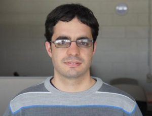
Guillermo Carbajal
Guillermo is on the teching faculty of Instituto de Ingeniería Eléctrica, Facultad de Ingeniería Universidad de la República in Montevideo Uruguay. He has been in the Perk Lab as visiting researcher and now is local champion of SlicerIGT and PLUS. We look foward to his return to the perk Lab!
Carbajal, Guillermo; Gómez, Álvaro; Fichtinger, Gabor; Ungi, Tamas
Portable Optically Tracked Ultrasound System for Scoliosis Measurement Conference
Recent Advances in Computational Methods and Clinical Applications for Spine Imaging (MICCAI 2014), vol. 20, Springer International Publishing Springer International Publishing, Cambridge, MA, 2015, ISBN: 978-3-319-14147-3.
@conference{Carbajal2015,

title = {Portable Optically Tracked Ultrasound System for Scoliosis Measurement},

author = {Guillermo Carbajal and Álvaro Gómez and Gabor Fichtinger and Tamas Ungi},

url = {http://link.springer.com/chapter/10.1007/978-3-319-14148-0_4

https://labs.cs.queensu.ca/perklab/wp-content/uploads/sites/3/2024/02/Carbajal2015.pdf},

doi = {10.1007/978-3-319-14148-0_4},

isbn = {978-3-319-14147-3},

year  = {2015},

date = {2015-02-01},

urldate = {2015-02-01},

booktitle = {Recent Advances in Computational Methods and Clinical Applications for Spine Imaging (MICCAI 2014)},

volume = {20},

pages = {37-46},

publisher = {Springer International Publishing},

address = {Cambridge, MA},

organization = {Springer International Publishing},

abstract = {<p><span style="font-family:helvetica neue,arial,helvetica,sans-serif">Monitoring spinal curvature in adolescent kyphoscoliosis requires regular radiographic examinations; however, the applied ionizing radiation increases the risk of cancer. Ultrasound imaging is favorable over X-ray because it does not emit ionizing radiation. It has been shown in the past that tracked ultrasound can be used to localize vertebral transverse processes as landmarks along the spine to measure curvature angles. Tests have been performed with spine phantoms, but scanning protocol, tracking system, data acquisition and processing time has not been considered in human subjects yet. In this paper, a portable optically tracked ultrasound system for scoliosis measurement is presented. It provides a simple way to acquire data in the clinical environment with the aim of comparing results to current X-ray-based measurement. The workflow of the procedure was tested on volunteers. The customized open-source software is shared with the community as part of our effort to make a clinically practical system.</span></p>},

keywords = {},

pubstate = {published},

tppubtype = {conference}

}
Carbajal, Guillermo; Gómez, Álvaro; Fichtinger, Gabor; Ungi, Tamas
Portable optically tracked ultrasound system for scoliosis measurement Journal Article
In: Recent Advances in Computational Methods and Clinical Applications for Spine Imaging, pp. 37-46, 2015.
@article{fichtinger2015k,

title = {Portable optically tracked ultrasound system for scoliosis measurement},

author = {Guillermo Carbajal and Álvaro Gómez and Gabor Fichtinger and Tamas Ungi},

url = {https://link.springer.com/chapter/10.1007/978-3-319-14148-0_4},

year  = {2015},

date = {2015-01-01},

journal = {Recent Advances in Computational Methods and Clinical Applications for Spine Imaging},

pages = {37-46},

publisher = {Springer International Publishing},

abstract = {Monitoring spinal curvature in adolescent kyphoscoliosis requires regular radiographic examinations; however, the applied ionizing radiation increases the risk of cancer. Ultrasound imaging is favorable over X-ray because it does not emit ionizing radiation. It has been shown in the past that tracked ultrasound can be used to localize vertebral transverse processes as landmarks along the spine to measure curvature angles. Tests have been performed with spine phantoms, but scanning protocol, tracking system, data acquisition and processing time has not been considered in human subjects yet. In this paper, a portable optically tracked ultrasound system for scoliosis measurement is presented. It provides a simple way to acquire data in the clinical environment with the aim of comparing results to current X-ray-based measurement. The workflow of the procedure was tested on volunteers. The customized …},

keywords = {},

pubstate = {published},

tppubtype = {article}

}
Carbajal, Guillermo; Lasso, Andras; Gómez, Álvaro; Fichtinger, Gabor
Improving N-Wire Phantom-based Freehand Ultrasound Calibration Conference
ImNO2013 - Imaging Network Ontario Symposium, vol. 8, 2013.
@conference{Carbajal2013c,

title = {Improving N-Wire Phantom-based Freehand Ultrasound Calibration},

author = {Guillermo Carbajal and Andras Lasso and Álvaro Gómez and Gabor Fichtinger},

url = {https://labs.cs.queensu.ca/perklab/wp-content/uploads/sites/3/2024/02/Carbajal2013b.pdf},

doi = {10.1007/s11548-013-0904-9},

year  = {2013},

date = {2013-11-01},

urldate = {2013-11-01},

booktitle = {ImNO2013 - Imaging Network Ontario Symposium},

journal = {IJCARS},

volume = {8},

pages = {1063-1072},

abstract = {<p>Purpose: Freehand tracked ultrasound imaging is an inexpensive non-invasive technique used in several guided interventions. This technique requires spatial calibration between the tracker and the ultrasound image plane. Several calibration devices (a.k.a. phantoms) use N-wires that are convenient for automatic procedures since the segmentation of fiducials in the images and the localization of the middle wires in space are straightforward and can be performed in real time. The procedures reported in literature consider only the spatial position of the middle wire. We investigate if better results can be achieved if the information of all the wires is equally taken into account. We also evaluated the precision and accuracy of the implemented methods to allow comparison with other methods. Methods: We consider a cost function based on the in-plane errors between the intersection of all the wires with the image plane and their respective segmented points in the image. This cost function is minimized iteratively starting from a seed computed with a closed-form solution based on the middle wires. Results: Mean calibration precision achieved with the N-wire phantom was about 0.5 mm using a shallow probe and mean accuracy was around 1.4 mm with all implemented methods. Precision was about 2.0 mm using a deep probe. Conclusions: Precision and accuracy achieved with the N-wire phantom and a shallow probe are at least comparable to that obtained with other methods traditionally considered more precise. Calibration using N-wires can be done more consistently if the parameters are optimized with the proposed 2D cost function.</p>},

keywords = {},

pubstate = {published},

tppubtype = {conference}

}
Carbajal, Guillermo; Lasso, Andras; Gómez, Álvaro; Fichtinger, Gabor
Improving N-wire phantom-based freehand ultrasound calibration Journal Article
In: International journal of computer assisted radiology and surgery, vol. 8, pp. 1063-1072, 2013.
@article{fichtinger2013b,

title = {Improving N-wire phantom-based freehand ultrasound calibration},

author = {Guillermo Carbajal and Andras Lasso and Álvaro Gómez and Gabor Fichtinger},

url = {https://link.springer.com/article/10.1007/s11548-013-0904-9},

year  = {2013},

date = {2013-01-01},

journal = {International journal of computer assisted radiology and surgery},

volume = {8},

pages = {1063-1072},

publisher = {Springer Berlin Heidelberg},

abstract = {Purpose

 Freehand tracked ultrasound imaging is an inexpensive non-invasive technique used in several guided interventions. This technique requires spatial calibration between the tracker and the ultrasound image plane. Several calibration devices (a.k.a. phantoms) use N-wires that are convenient for automatic procedures since the segmentation of fiducials in the images and the localization of the middle wires in space are straightforward and can be performed in real time. The procedures reported in literature consider only the spatial position of the middle wire. We investigate if better results can be achieved if the information of all the wires is equally taken into account. We also evaluated the precision and accuracy of the implemented methods to allow comparison with other methods.

Methods

 We consider a cost function based on the in-plane …},

keywords = {},

pubstate = {published},

tppubtype = {article}

}
Freehand tracked ultrasound imaging is an inexpensive non-invasive technique used in several guided interventions. This technique requires spatial calibration between the tracker and the ultrasound image plane. Several calibration devices (a.k.a. phantoms) use N-wires that are convenient for automatic procedures since the segmentation of fiducials in the images and the localization of the middle wires in space are straightforward and can be performed in real time. The procedures reported in literature consider only the spatial position of the middle wire. We investigate if better results can be achieved if the information of all the wires is equally taken into account. We also evaluated the precision and accuracy of the implemented methods to allow comparison with other methods.
Methods
We consider a cost function based on the in-plane …