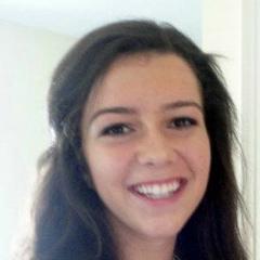
Maggie Hess
During the summer in 2014, Maggie worked on quantitative MR image-based analysis of clot lysis in the brain induced by focused ultrasound ablation, in collabporation between the Hospital for Sick Kids and the Perk Lab, she was deployed at Sick Kids working with Dr. James Drake in pediatric neurosurgery. She will be back in the Perk Lab since September, as undergraduate research assistant, working in various projects where she develops image processing workflows (segmentation, registration, meshing) in 3D Slicer for various image-guided surgery applications. Maggie graduated from Queen's in 2015 and continues her studies in medical school at the University of Toronto.
Hess, Maggie; Eastwood, Kyle; Linder, Bence; Bodani, Vivek; Lasso, Andras; Looi, Thomas; Fichtinger, Gabor; Drake, James
Visual design and verification tool for collision-free dexterous patient specific neurosurgical instruments Journal Article
In: vol. 9786, pp. 486-503, 2016.
@article{fichtinger2016i,

title = {Visual design and verification tool for collision-free dexterous patient specific neurosurgical instruments},

author = {Maggie Hess and Kyle Eastwood and Bence Linder and Vivek Bodani and Andras Lasso and Thomas Looi and Gabor Fichtinger and James Drake},

url = {https://www.spiedigitallibrary.org/conference-proceedings-of-spie/9786/97861M/Visual-design-and-verification-tool-for-collision-free-dexterous-patient/10.1117/12.2217304.short},

year  = {2016},

date = {2016-01-01},

volume = {9786},

pages = {486-503},

publisher = {SPIE},

abstract = {PURPOSE: In many minimally invasive neurosurgical procedures, the surgical workspace is a small tortuous cavity that is accessed using straight, rigid instruments with limited dexterity. Specifically considering neuroendoscopy, it is often challenging for surgeons, using standard instruments, to reach multiple surgical targets from a single incision. To address this problem, continuum tools are under development to create highly dexterous minimally invasive instruments. However, this design process is not trivial, and therefore, a user-friendly design platform capable of easily incorporating surgeon input is needed.

METHODS: We propose a method that uses simulation and visual verification to design continuum tools that are patient and procedure specific. Our software module utilizes pre-operative scans and virtual threedimensional (3D) patient models to intuitively aid instrument design. The user specifies basic …},

keywords = {},

pubstate = {published},

tppubtype = {article}

}
METHODS: We propose a method that uses simulation and visual verification to design continuum tools that are patient and procedure specific. Our software module utilizes pre-operative scans and virtual threedimensional (3D) patient models to intuitively aid instrument design. The user specifies basic …
Hess, Maggie; Drake, James
Quantification of intraventricular blood clot in MR-guided focused ultrasound surgery Conference
SPIE Medical Imaging 2015: Image-Guided Procedures, Robotic Interventions, and Modeling, vol. 9415, Orlando, Florida, 2015.
@conference{Hess2014a,

title = {Quantification of intraventricular blood clot in MR-guided focused ultrasound surgery},

author = {Maggie Hess and James Drake},

editor = {Thomas Looi and Andras Lasso and Gabor Fichtinger},

url = {http://proceedings.spiedigitallibrary.org/proceeding.aspx?articleid=2210357

https://labs.cs.queensu.ca/perklab/wp-content/uploads/sites/3/2024/02/Hess2014a-manuscript_0.pdf},

doi = {10.1117/12.2081330},

year  = {2015},

date = {2015-03-01},

urldate = {2015-03-01},

booktitle = {SPIE Medical Imaging 2015: Image-Guided Procedures, Robotic Interventions, and Modeling},

volume = {9415},

pages = {94152J1-94152J9},

address = {Orlando, Florida},

abstract = {<div class="page" title="Page 1"> <div class="layoutArea"> <div class="column"> <p><strong>Purpose: </strong><span style="font-family:timesnewromanpsmt; font-size:10.000000pt">Intraventricular hemorrhage (IVH) affects nearly 15% of preterm infants. It can lead to ventricular dilation and cognitive impairment. To ablate IVH clots, MR-guided focused ultrasound surgery (MRgFUS) is investigated. This procedure requires accurate, fast and consistent quantification of ventricle and clot volumes.</span><br /> <strong>Methods</strong><span style="font-family:timesnewromanpsmt; font-size:10.000000pt">: We developed a semi-autonomous segmentation (SAS) algorithm for measuring changes in the ventricle and clot volumes. Images are normalized, and then ventricle and clot masks are registered to the images. Voxels of the registered masks and voxels obtained by thresholding the normalized images are used as seed points for competitive region growing, which provides the final segmentation. The user selects the areas of interest for correspondence after thresholding and these selections are the final seeds for region growing. SAS was evaluated on an IVH porcine model. </span><strong>Results</strong><span style="font-family:timesnewromanpsmt; font-size:10.000000pt">: SAS was compared to ground truth manual segmentation (MS) for accuracy, efficiency, and consistency. Accuracy was determined by comparing clot and ventricle volumes produced by SAS and MS, and comparing contours by calculating 95% Hausdorff distances between the two labels. In Two-One-Sided Test, SAS and MS were found to be significantly equivalent (p < 0.01). SAS on average was found to be 15 times faster than MS (p < 0.01). Consistency was determined by repeated segmentation of the same image by both SAS and manual methods, SAS being significantly more consistent than MS (p < 0.05). </span></p> <p><strong>Conclusion</strong><span style="font-family:timesnewromanpsmt; font-size:10.000000pt">: SAS is a viable method to quantify the IVH clot and the lateral brain ventricles and it is serving in a large- scale porcine study of MRgFUS treatment of IVH clot lysis. </span></p>

</div>

</div>

</div>},

keywords = {},

pubstate = {published},

tppubtype = {conference}

}
</div>
</div>
</div>