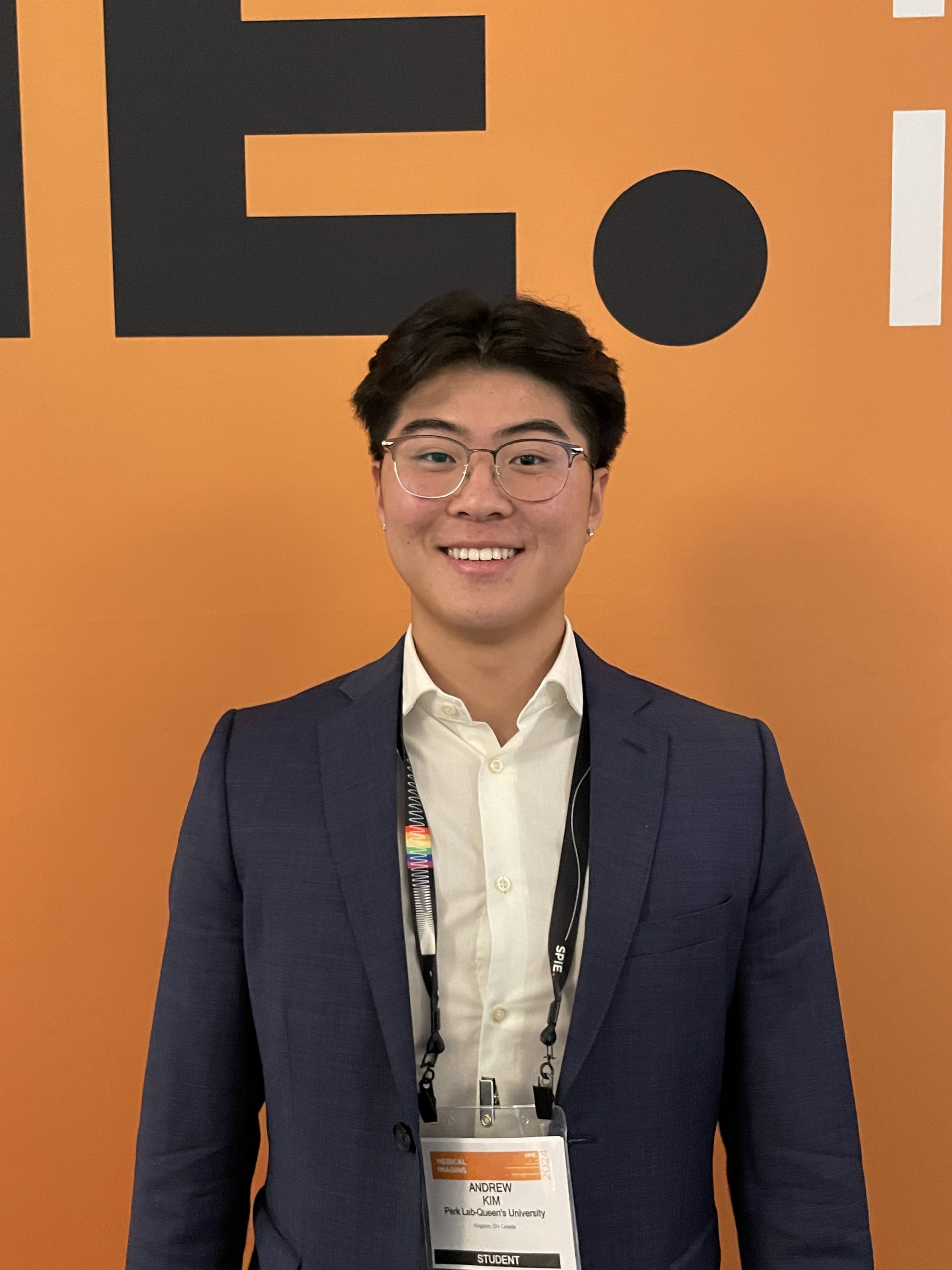
Andrew Kim
Undergraduate Student
School of Computing
Queen’s University
Member from January 2023 to Current
Biography
Andrew is an undergraduate student in the Biomedical Computing program. Research interests include surgical navigation and applications of deep learning for surgery.
Publications
Kim, Andrew S.; Yeung, Chris; Szabo, Robert; Sunderland, Kyle; Hisey, Rebecca; Morton, David; Kikinis, Ron; Diao, Babacar; Mousavi, Parvin; Ungi, Tamas; Fichtinger, Gabor
SPIE, 2024.
@proceedings{Kim2024,

title = {Percutaneous nephrostomy needle guidance using real-time 3D anatomical visualization with live ultrasound segmentation},

author = {Andrew S. Kim and Chris Yeung and Robert Szabo and Kyle Sunderland and Rebecca Hisey and David Morton and Ron Kikinis and Babacar Diao and Parvin Mousavi and Tamas Ungi and Gabor Fichtinger},

editor = {Maryam E. Rettmann and Jeffrey H. Siewerdsen},

doi = {10.1117/12.3006533},

year  = {2024},

date = {2024-03-29},

urldate = {2024-03-29},

publisher = {SPIE},

abstract = {

PURPOSE: Percutaneous nephrostomy is a commonly performed procedure to drain urine to provide relief in patients with hydronephrosis. Conventional percutaneous nephrostomy needle guidance methods can be difficult, expensive, or not portable. We propose an open-source real-time 3D anatomical visualization aid for needle guidance with live ultrasound segmentation and 3D volume reconstruction using free, open-source software. METHODS: Basic hydronephrotic kidney phantoms were created, and recordings of these models were manually segmented and used to train a deep learning model that makes live segmentation predictions to perform live 3D volume reconstruction of the fluid-filled cavity. Participants performed 5 needle insertions with the visualization aid and 5 insertions with ultrasound needle guidance on a kidney phantom in randomized order, and these were recorded. Recordings of the trials were analyzed for needle tip distance to the center of the target calyx, needle insertion time, and success rate. Participants also completed a survey on their experience. RESULTS: Using the visualization aid showed significantly higher accuracy, while needle insertion time and success rate were not statistically significant at our sample size. Participants mostly responded positively to the visualization aid, and 80% found it easier to use than ultrasound needle guidance. CONCLUSION: We found that our visualization aid produced increased accuracy and an overall positive experience. We demonstrated that our system is functional and stable and believe that the workflow with this system can be applied to other procedures. This visualization aid system is effective on phantoms and is ready for translation with clinical data.},

keywords = {},

pubstate = {published},

tppubtype = {proceedings}

}
PURPOSE: Percutaneous nephrostomy is a commonly performed procedure to drain urine to provide relief in patients with hydronephrosis. Conventional percutaneous nephrostomy needle guidance methods can be difficult, expensive, or not portable. We propose an open-source real-time 3D anatomical visualization aid for needle guidance with live ultrasound segmentation and 3D volume reconstruction using free, open-source software. METHODS: Basic hydronephrotic kidney phantoms were created, and recordings of these models were manually segmented and used to train a deep learning model that makes live segmentation predictions to perform live 3D volume reconstruction of the fluid-filled cavity. Participants performed 5 needle insertions with the visualization aid and 5 insertions with ultrasound needle guidance on a kidney phantom in randomized order, and these were recorded. Recordings of the trials were analyzed for needle tip distance to the center of the target calyx, needle insertion time, and success rate. Participants also completed a survey on their experience. RESULTS: Using the visualization aid showed significantly higher accuracy, while needle insertion time and success rate were not statistically significant at our sample size. Participants mostly responded positively to the visualization aid, and 80% found it easier to use than ultrasound needle guidance. CONCLUSION: We found that our visualization aid produced increased accuracy and an overall positive experience. We demonstrated that our system is functional and stable and believe that the workflow with this system can be applied to other procedures. This visualization aid system is effective on phantoms and is ready for translation with clinical data.