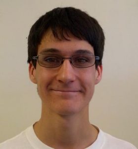
Ian Cumming
Ian was a summer intern in the Perk Lab in 2013; he worked on designing patient-specific nedle catheter guides for high dose rate brachytherapy of superficial cancers.
Schumacher, Mark; Lasso, Andras; Cumming, Ian; Rankin, Adam; Falkson, Conrad; Schreiner, John; Joshi, C. P.; Fichtinger, Gabor
3D-printed surface mould applicator for high-dose-rate brachytherapy Conference
SPIE Medical Imaging 2015, vol. 9415, 2015.
@conference{Schumacher2015,

title = {3D-printed surface mould applicator for high-dose-rate brachytherapy},

author = {Mark Schumacher and Andras Lasso and Ian Cumming and Adam Rankin and Conrad Falkson and John Schreiner and C. P. Joshi and Gabor Fichtinger},

url = {https://labs.cs.queensu.ca/perklab/wp-content/uploads/sites/3/2024/02/Schumacher2015-manuscript.pdf

https://labs.cs.queensu.ca/perklab/wp-content/uploads/sites/3/2024/02/Schumacher2015-poster.pdf},

year  = {2015},

date = {2015-01-01},

urldate = {2015-01-01},

booktitle = {SPIE Medical Imaging 2015},

volume = {9415},

abstract = {<p><strong>ABSTRACT</strong></p>

<p>PURPOSE: In contemporary high-dose-rate brachytherapy treatment of superficial tumors, catheters are placed in a wax mould. The creation of current wax models is a difficult and time consuming proces.The irradiation plan can only be computed post-construction and requires a second CT scan. In case no satisfactory dose plan can be created, the mould is discarded and the process is repeated. The objective of this work was to develop an automated method to replace suboptimal wax moulding. METHODS: We developed a method to design and manufacture moulds that guarantee to yield satisfactory dosimetry. A 3D-printed mould with channels for the catheters designed from the patient’s CT and mounted on a patient-specific thermoplastic mesh mask. The mould planner was implemented as an open-source module in the 3D Slicer platform. RESULTS: Series of test moulds were created to accommodate standard brachytherapy catheters of 1.70mm diameter. A calibration object was used to conclude that tunnels with a diameter of 2.25mm, minimum 12mm radius of curvature, and 1.0mm open channel gave the best fit for this printer/catheter combination. Moulds were created from the CT scan of thermoplastic mesh masks of actual patients. The patient-specific moulds have been visually verified to fit on the thermoplastic meshes. CONCLUSION: The masks were visually shown to fit onto the thermoplastic meshes, next the resulting dosimetry will have to be compared with treatment plans and dosimetry achieved with conventional wax moulds in order to validate our 3D printed moulds.</p>},

keywords = {},

pubstate = {published},

tppubtype = {conference}

}
<p>PURPOSE: In contemporary high-dose-rate brachytherapy treatment of superficial tumors, catheters are placed in a wax mould. The creation of current wax models is a difficult and time consuming proces.The irradiation plan can only be computed post-construction and requires a second CT scan. In case no satisfactory dose plan can be created, the mould is discarded and the process is repeated. The objective of this work was to develop an automated method to replace suboptimal wax moulding. METHODS: We developed a method to design and manufacture moulds that guarantee to yield satisfactory dosimetry. A 3D-printed mould with channels for the catheters designed from the patient’s CT and mounted on a patient-specific thermoplastic mesh mask. The mould planner was implemented as an open-source module in the 3D Slicer platform. RESULTS: Series of test moulds were created to accommodate standard brachytherapy catheters of 1.70mm diameter. A calibration object was used to conclude that tunnels with a diameter of 2.25mm, minimum 12mm radius of curvature, and 1.0mm open channel gave the best fit for this printer/catheter combination. Moulds were created from the CT scan of thermoplastic mesh masks of actual patients. The patient-specific moulds have been visually verified to fit on the thermoplastic meshes. CONCLUSION: The masks were visually shown to fit onto the thermoplastic meshes, next the resulting dosimetry will have to be compared with treatment plans and dosimetry achieved with conventional wax moulds in order to validate our 3D printed moulds.</p>
Cumming, Ian; Joshi, C. P.; Lasso, Andras; Rankin, Adam; Falkson, Conrad; Schreiner, John; Fichtinger, Gabor
American Association Physicists in Medicine (AAPM), vol. 41 (abstract in Medical Physics), no. 222, 2014.
@conference{Cumming2014,

title = {3D Printed Patient-Specific Surface Mould Applicators for Brachytherapy Treatment of Superficial Lesions},

author = {Ian Cumming and C. P. Joshi and Andras Lasso and Adam Rankin and Conrad Falkson and John Schreiner and Gabor Fichtinger},

url = {https://labs.cs.queensu.ca/perklab/wp-content/uploads/sites/3/2024/02/Cumming2014.pdf},

doi = {http://dx.doi.org/10.1118/1.4888333},

year  = {2014},

date = {2014-06-01},

urldate = {2014-06-01},

booktitle = {American Association Physicists in Medicine (AAPM)},

volume = {41 (abstract in Medical Physics)},

number = {222},

abstract = {<p>Purpose:<br />

Evaluate the feasibility of constructing 3D-printed patient-specific surface mould applicators for HDR brachytherapy treatment of superficial lesions.<br />

<br />

Methods:<br />

We propose using computer-aided design software to create 3D printed surface mould applicators for brachytherapy. A mould generation module was developed in the open-source 3D Slicer (www.slicer.org) medical image analysis platform. The system extracts the skin surface from CT images, and generates smooth catheter paths over the region of interest based on user-defined start and end points at a specified stand-off distance from the skin surface. The catheter paths are radially extended to create catheter channels that are sufficiently wide to ensure smooth insertion of catheters for a safe source travel. An outer mould surface is generated to encompass the channels. The mould is also equipped with fiducial markers to ensure its reproducible placement.<br />

<br />

A surface mould applicator with eight parallel catheter channels of 4mm diameters was fabricated for the nose region of a head phantom; flexible plastic catheters of 2mm diameter were threaded through these channels maintaining 10mm catheter separations and a 5mm stand-off distance from the skin surface. The apparatus yielded 3mm thickness of mould material between channels and the skin. The mould design was exported as a stereolithography file to a Dimension SST1200es 3D printer and printed using ABS Plus plastic material.<br />

<br />

Results:<br />

The applicator closely matched its design and was found to be sufficiently rigid without deformation during repeated application on the head phantom. Catheters were easily threaded into channels carved along catheter paths. Further tests are required to evaluate feasibility of channel diameters smaller than 4mm.<br />

<br />

Conclusion:<br />

Construction of 3D-printed mould applicators show promise for use in patient specific brachytherapy of superficial lesions. Further evaluation of 3D printing techniques and materials is required for constructing sufficiently thin, rigid and durable surface moulds suitable for clinical deployment.<br />

 </p>},

keywords = {},

pubstate = {published},

tppubtype = {conference}

}
Evaluate the feasibility of constructing 3D-printed patient-specific surface mould applicators for HDR brachytherapy treatment of superficial lesions.<br />
<br />
Methods:<br />
We propose using computer-aided design software to create 3D printed surface mould applicators for brachytherapy. A mould generation module was developed in the open-source 3D Slicer (www.slicer.org) medical image analysis platform. The system extracts the skin surface from CT images, and generates smooth catheter paths over the region of interest based on user-defined start and end points at a specified stand-off distance from the skin surface. The catheter paths are radially extended to create catheter channels that are sufficiently wide to ensure smooth insertion of catheters for a safe source travel. An outer mould surface is generated to encompass the channels. The mould is also equipped with fiducial markers to ensure its reproducible placement.<br />
<br />
A surface mould applicator with eight parallel catheter channels of 4mm diameters was fabricated for the nose region of a head phantom; flexible plastic catheters of 2mm diameter were threaded through these channels maintaining 10mm catheter separations and a 5mm stand-off distance from the skin surface. The apparatus yielded 3mm thickness of mould material between channels and the skin. The mould design was exported as a stereolithography file to a Dimension SST1200es 3D printer and printed using ABS Plus plastic material.<br />
<br />
Results:<br />
The applicator closely matched its design and was found to be sufficiently rigid without deformation during repeated application on the head phantom. Catheters were easily threaded into channels carved along catheter paths. Further tests are required to evaluate feasibility of channel diameters smaller than 4mm.<br />
<br />
Conclusion:<br />
Construction of 3D-printed mould applicators show promise for use in patient specific brachytherapy of superficial lesions. Further evaluation of 3D printing techniques and materials is required for constructing sufficiently thin, rigid and durable surface moulds suitable for clinical deployment.<br />
</p>
Cumming, Ian; Joshi, Chandra; Lasso, Andras; Rankin, Adam; Falkson, Conrad; Schreiner, L John; Fichtinger, Gabor
SU-ET-04: 3D printed patient-specific surface mould applicators for brachytherapy treatment of superficial lesions Journal Article
In: Med Phys, vol. 41, iss. 6 part 11, pp. 222, 2014.
@article{fichtinger2014k,

title = {SU-ET-04: 3D printed patient-specific surface mould applicators for brachytherapy treatment of superficial lesions},

author = {Ian Cumming and Chandra Joshi and Andras Lasso and Adam Rankin and Conrad Falkson and L John Schreiner and Gabor Fichtinger},

url = {http://perk.cs.queensu.ca/sites/perkd7.cs.queensu.ca/files/Cumming2014.pdf},

year  = {2014},

date = {2014-01-01},

journal = {Med Phys},

volume = {41},

issue = {6 part 11},

pages = {222},

abstract = {Purpose: Evaluate the feasibility of constructing 3D-printed patient-specific surface mould applicators for HDR brachytherapy treatment of superficial lesions.

Methods: We propose using computer-aided design software to create 3D-printed surface mould applicators for brachytherapy. A mould generation module was developed in the opensource 3D Slicer (www. slicer. org) medical image analysis platform. The system extracts the skin surface from CT images, and generates smooth catheter paths over the region of interest based on user-defined start and end points at a specified stand-off distance from the skin surface. The catheter paths are radially extended to create catheter channels that are sufficiently wide to ensure smooth insertion of catheters for a safe source travel. An outer mould surface is generated to encompass the channels. The mould is also equipped with fiducial markers to ensure its reproducible placement.},

keywords = {},

pubstate = {published},

tppubtype = {article}

}
Methods: We propose using computer-aided design software to create 3D-printed surface mould applicators for brachytherapy. A mould generation module was developed in the opensource 3D Slicer (www. slicer. org) medical image analysis platform. The system extracts the skin surface from CT images, and generates smooth catheter paths over the region of interest based on user-defined start and end points at a specified stand-off distance from the skin surface. The catheter paths are radially extended to create catheter channels that are sufficiently wide to ensure smooth insertion of catheters for a safe source travel. An outer mould surface is generated to encompass the channels. The mould is also equipped with fiducial markers to ensure its reproducible placement.